Balloon Mitral Valvotomy
Balloon Mitral Valvotomy | Cardiology | Apex Hospitals

What is Balloon Mitral Valvotomy (BMV)?
Balloon mitral valvotomy (BMV) is a medical procedure used to treat mitral stenosis, a condition where the mitral valve in the heart becomes narrowed. The mitral valve regulates the flow of blood between the left atrium and the left ventricle. When this valve becomes stenotic, it restricts the normal flow of blood from the left atrium to the left ventricle, hindering the efficient functioning of the heart.
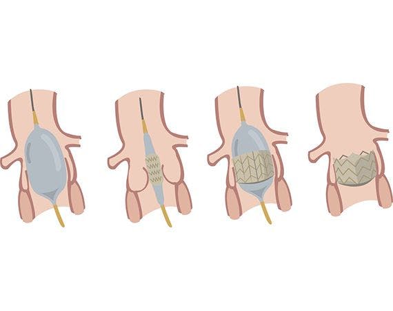
During a balloon mitral valvotomy, a catheter with a deflated balloon at its tip is inserted into a blood vessel, typically the femoral vein, and threaded up to the heart. Once the catheter is in position, the balloon is inflated, which opens and widens the narrowed mitral valve. This process helps improve blood flow, reducing the symptoms associated with mitral stenosis, such as fatigue, shortness of breath, and chest discomfort.
Balloon mitral valvotomy is considered a less invasive alternative to open-heart surgery for selected patients with mitral stenosis. It is particularly effective in cases where the mitral valve leaflets are pliable and suitable for the balloon dilation technique. The procedure is often performed by interventional cardiologists with expertise in catheter-based interventions. It can lead to symptom relief and improved quality of life for individuals with mitral stenosis.
Who needs Balloon mitral valvotomy (BMV)?
Balloon mitral valvotomy emerges as a potential treatment for individuals experiencing symptoms stemming from various heart conditions:
Aortic Valve Stenosis
The aortic valve regulates blood flow between the lower left heart chamber (ventricle) and the body's primary pumping artery (aorta). In cases of aortic stenosis, where the valve narrows, hindering proper blood flow through the heart, our cardiologists may recommend valvuloplasty. This option is particularly considered for patients who are not suitable candidates for aortic valve repair, replacement surgery, or transcatheter aortic valve replacement (TAVR). In some instances, the aortic valve may narrow again after valvuloplasty, prompting cardiologists to use it as a temporary treatment before TAVR or surgical valve replacement.
Mitral Stenosis
The mitral valve manages blood flow between the upper left heart chamber (atrium) and the left ventricle. Balloon mitral valvotomy is often the preferred treatment for mitral valve stenosis. Additionally, our cardiologists may explore mitral valve repair, replacement surgery, and transcatheter mitral valve replacement (TMVR) based on individual cases.
Pulmonary Stenosis
Controlling blood flow from the right ventricle to the lungs, the pulmonary valve may experience stenosis, often a congenital condition addressed during childhood. Adult patients may require further treatment over time. Our cardiologists may employ valvuloplasty, pulmonary valve repair, replacement surgery, or transcatheter pulmonary valve replacement (TPVR) to manage pulmonary stenosis. The choice of treatment depends on individual circumstances and the severity of the condition.
What to Anticipate During a Balloon mitral valvotomy Procedure?
Balloon valvuloplasty is an inpatient procedure with an approximate duration of one hour, ensuring your comfort through administering anaesthesia. Our collaborative team comprises an interventional cardiologist, an interventional echocardiographer, and an anaesthesia specialist, collectively executing the procedure.
As the procedure begins, the team attaches you to an electrocardiogram machine, monitoring your heart's electrical activity. Additionally, various machines record vital signs, including heart rate, breathing, oxygen levels, and blood pressure.
Throughout the balloon valvotomy, the interventional cardiologist follows these steps:
1. Introduce a hollow plastic tube, known as a sheath, through a blood vessel in your groin.
2. Inserts a catheter, featuring a balloon at the tip, through the sheath.
3. Guides the catheter to your heart, utilizing X-ray and echocardiographic images to visualise heart structures and blood vessels precisely.
4. A contrast dye is administered through the catheter, enhancing the visibility of the narrowed heart valve.
5. Position the balloon within the narrowed heart valve, occasionally requesting you to hold your breath for a few seconds. The balloon may need inflation and deflation cycles to open the valve fully.
6. Inflate the balloon to expand the valve opening.
7. Upon successfully opening the valve, the interventional cardiologist removed the catheter and deflated the balloon.
This comprehensive process ensures the adequate dilation of the heart valve, and the removal of the catheter marks the completion of the valvuloplasty procedure.
Speak to our team about Balloon Mitral Valvotomy
If you're considering or have questions about balloon mitral valvotomy, our dedicated team is here to provide the information and support you need.
Our team, consisting of experienced cardiologists and healthcare professionals, is ready to discuss the specifics of balloon mitral valvotomy, including its benefits, the procedure itself, and what you can expect regarding recovery. Whether you are exploring treatment options, seeking clarification, or preparing for the process, our experts are committed to guiding you through every step.
Feel free to reach out to schedule a consultation or discuss any questions you may have. Your heart health is our priority, and we are here to provide personalized information and support tailored to your individual needs. Contact us today to speak with our knowledgeable team about balloon mitral valvotomy and to address any concerns you may have.
FAQS
Health In A Snap, Just One App.
KNOW MORE
